
2026-01-02 Posted by TideChem view:467
Bioconjugation is key to many new ways to treat diseases, diagnose problems, and engineer proteins. The right choice of a PEG linker can make or break your experiment, even if the rest of your process is solid.
If your PEG linker isn't a good fit, you might not get enough of your product, it could clump up, lose its activity, or act unpredictably in the body. But a good PEG linker can increase how well it dissolves, make it more stable, and change how it acts in the body—all without stopping it from binding to its target or doing what it's supposed to.
If you're new to PEG linkers, check out our overview article: What are PEG Linkers? This guide will show you how to choose the best PEG linker for bioconjugation, based on what you actually need rather than just general info.
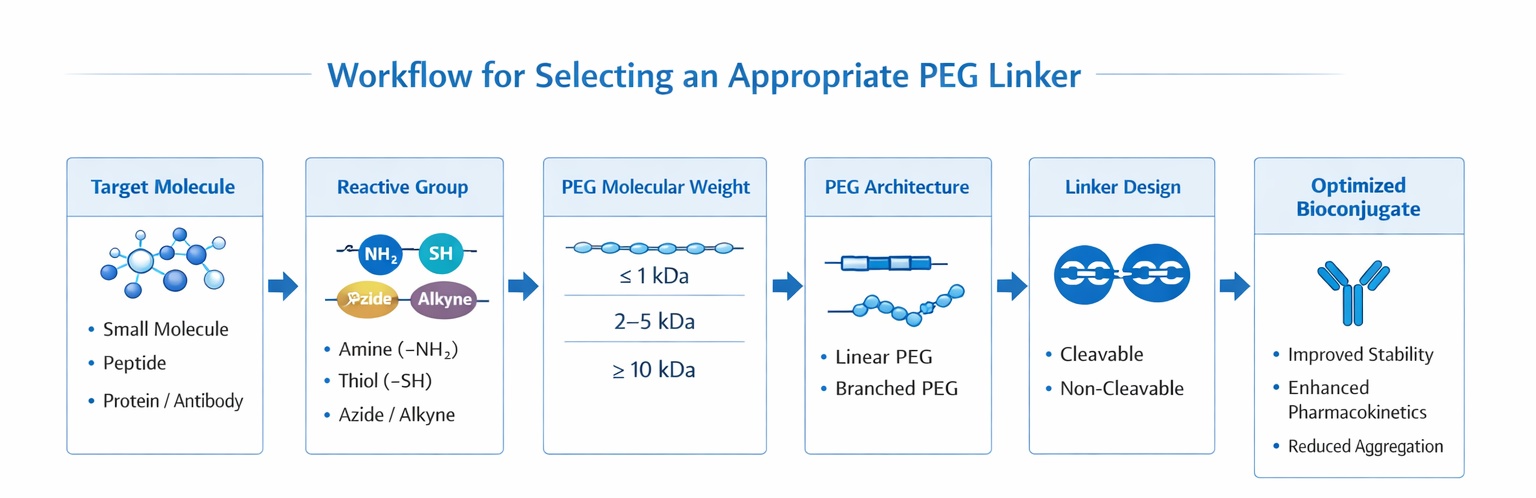
Workflow for Selecting an Appropriate PEG Linker
Picking a PEG linker means thinking about lots of things. What's best depends on the chemistry you're using, the shape of the PEG, its size, and whether it needs to break apart inside the body. Think about all these things together.
The ends of a PEG linker decide what it can react with, how well it reacts with only that, and when the conjugation happens. So, always think about whether the functional groups are a match first.
Different PEG linkers are made to react with certain parts of biomolecules—like amines, thiols, carboxyl groups, or azides/alkynes. If the chemistry doesn't match, you'll probably end up with not enough of your product or a mix of different things.
| PEG Linker Functional Group | Target Functional Group on Biomolecule | Typical Reaction Conditions | Common Applications |
| NHS ester | Primary amines (Lys, N-terminus) | pH 7.5–8.5, aqueous buffer | Protein PEGylation, ADCs |
| Maleimide | Thiols (Cys) | pH 6.5–7.5 | Site-specific protein conjugation |
| Azide | Alkyne (click chemistry) | CuAAC or SPAAC | Bioorthogonal labeling |
| Alkyne | Azide (click chemistry) | CuAAC or SPAAC | Imaging probes, bioconjugation |
| Hydrazide | Aldehyde / ketone | Mild acidic conditions | Glycoprotein conjugation |
| Carboxyl (–COOH) | Amine (via EDC/NHS) | pH 4.5–7.5 | General-purpose conjugation |
Table: Common PEG linker functional groups, target residues, reaction conditions, and typical applications
When picking a functional group, think not just about how well it reacts but also the reaction's pH, how stable it is when stored, and if it might react with other things you don't want it to.
For a broader understanding of how different functionalized PEG linkers are designed and applied across bioconjugation strategies, see our comprehensive overview on What are PEG Linkers?
The molecular weight of PEG greatly affects how well it dissolves, how flexible it is, how well it hides from the body, and how long it lasts in the blood. But bigger isn't always better. Too much PEG can get in the way of binding or lower its activity.
| PEG Molecular Weight | Key Characteristics | Advantages | Typical Use Cases |
| < 2 kDa | Short, limited shielding | Minimal impact on activity | Small-molecule conjugation |
| 2–5 kDa | Moderate flexibility | Improved solubility without bulk | Imaging probes, peptide labeling |
| 5–10 kDa | Balanced shielding | Good PK improvement | Peptides, proteins, ADCs |
| 10–20 kDa | Strong steric protection | Extended circulation time | Therapeutic proteins |
| > 20 kDa | Very high shielding | Maximum half-life extension | Long-acting biologics (with caution) |
Table: PEG molecular weight ranges and recommended use cases
The shape of the PEG matters a lot but often gets missed.
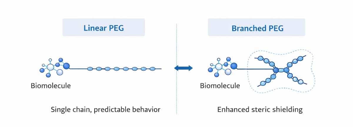
Diagram: Linear vs. branched PEG structures and steric shielding effect
Branched PEG linkers are often used in antibody–drug conjugates (ADCs) and attaching PEG to cytokines. For example, attaching branched PEG to interferon-alpha has been shown to make it last longer in the blood while keeping its activity higher than if you used linear PEG.
Another thing to think about is whether the PEG linker should stay put or let go of its cargo in the body.
The choice depends on whether you need the cargo to be released to work or whether you just need it to stay together.
| Application | Preferred PEG Type | Recommended MW Range | Key Selection Rationale |
| ADCs | Branched PEG | 5–10 kDa | Reduce aggregation, control DAR |
| Peptide conjugation | Linear PEG | 2–10 kDa | Preserve activity, improve stability |
| Protein PEGylation | Linear or branched PEG | 5–20 kDa | Extend half-life, reduce clearance |
| Diagnostics & imaging | Linear PEG | 1–5 kDa | Minimize steric interference |
| Targeted drug delivery | Cleavable PEG | Application-dependent | Controlled payload release |
Application-based PEG linker selection matrix
This part turns technical stuff into advice based on what you're using it for.
How well an ADC works really depends on the linker. PEG linkers affect how many drugs are on each antibody (DAR), how well it dissolves, how likely it is to clump, and how safe it is overall.
Branched PEG linkers are often better for ADCs because they hide it better, which stops clumping even when there are more drugs on each antibody. Mid-range branched PEGs (around 5–10 kDa) usually give you the best mix of flexibility, solubility, and control over how long it lasts in the body, without getting in the way of it binding to its target.
Peptides and proteins tend to break down quickly, clump up, and get cleared out fast. PEG linkers help with these problems by making them dissolve better in water, last longer in the blood, and not be recognized by the immune system.
For most peptide conjugations, linear PEGs in the 2–10 kDa range are good enough to make them more stable while keeping their activity. Bigger proteins or things that are easily messed up might do better with branched PEGs, which protect them more without changing their shape too much.
When you're diagnosing or imaging, it's really important to get a clear signal. PEG linkers make probes work better by stopping them from binding to the wrong things, helping them dissolve, and keeping fluorescent or radioactive stuff stable.
Linear PEGs in the 1–5 kDa range are often used for fluorescent labeling and radioimmunoconjugates because they make the probe act better without making it too bulky.
Avoiding these mistakes can really help your project:
Picking a PEG linker is complicated, and there's no single right answer. The chemistry, size, shape, and whether it breaks down need to match what you're trying to do.
Ultimately, successful bioconjugation starts with a solid understanding of PEG linker fundamentals. Our article What Are PEG Linkers? provides a comprehensive overview of PEG linker structures, properties, and application scenarios.
If you understand what each of these things does and match them to what you need, you can get more of your product, make it more stable, and make it work better overall. For hard or big projects, working with a PEG linker supplier that knows what they're doing can get you custom solutions and make it easier to produce large amounts.